


Data and Codes
BCI-VR Data : scalp-recorded EEG data of the brain-computer interface (BCI) in a virtual reality (VR) experiment
- BCI-VR data were recorded during neurorehabilitation training of post-stroke subjects in VR.
- Resting state EEG with the eyes-closed and eyes-open conditions was recorded before and after each training session.
- Each BCI-VR training session consists of three blocks of training, each with ten motor imagery (MI) trials.
- To discriminate between resting state and MI periods, we used time-scores of a PARAFAC atom, see our paper.
- Preliminary analysis was presented the the 17th International Work-Conference on Artificial Neural Networks, (IWANN2023), see our talk.
- Description of the experiment and EEG data technicalities can be downloaded here as a pdf file. Please contact us if further information is needed.
- A 66-year-old man with residual right-sided hemiparesis after a stroke.
- Data were recorded from October 13, 2022, to December 16, 2022, representing 16 training days.
- BCI-VR Data
- MATLAB files (zip file, 158 MB)
- EDF files (zip file, 60 MB)
- Resting state EC and EO Data
- MATLAB files (zip file, 56 MB)
- EDF files (zip file, 21 MB)
- An 84-year-old man with residual left-sided hemiparesis after a stroke.
- Data were recorded from May 11, 2023, to June 22, 2023, representing 13 training days.
- Before the training, after each training block, and after the training, a collection of questions focused on mental state and cybersickness was collected. The Excel document with the answers is linked here.
- BCI-VR Data
- MATLAB files (zip file, 119 MB)
- EDF files (zip file, 48 MB)
- Resting state EC and EO Data
- MATLAB files (zip file, 48 MB)
- EDF files (zip file, 18 MB)
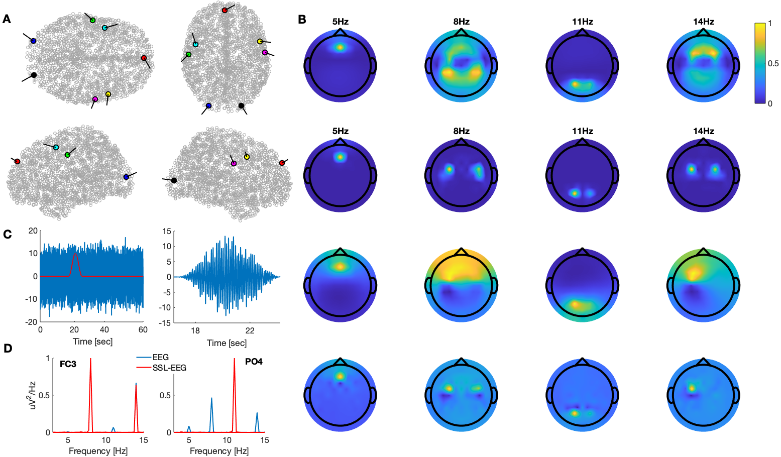
NBO Data (zip file, 46 MB): simulated narrow band scalp-recorded EEG oscillations
- We applied an anatomical forward model consisting of 2,004 dipoles placed in gray matter. This model was obtained from the on-line
repository http://mikexcohen.com/data/ (downloaded, June 20, 2020).
We modeled narrowband oscillatory sources with seven dipoles at four different frequencies- θ rhythm at 5 Hz, a dipole located at the frontal midline region
- μ rhythm at 8 Hz, left and right hemisphere dipoles located within the somatosensory cortex
- β rhythm at 14 Hz, left and right hemisphere dipoles located within the somatomotor cortex
- α rhythm at 11 Hz, left and right hemisphere dipoles located within the posterior cortex
- More details:
Rosipal R., Rostakova Z., Trejo L.J.
Tensor Decomposition of Human Narrowband Oscillatory Brain Activity in Frequency, Space and Time.
Biological Psychology, 169, 108287, 2022.
abstract
- If you are interested in any dataset published in our studies, please contact us.
- Kernel-based methods - kernel PLS, kernel SIMPLS, kernel PCR, kernel RR, EM kernel PCA
please send an e-mail to roman.rosipal@savba.sk